The Glymphatic System
Meets
The Cranial Rhythmic Impulse (CRI)
Do you have a loved one suffering from Alzheimer’s, Parkinson’s, Schizophrenia or ALS?
The glymphatic system, activated only during sleep, has potential significance for neurodegenerative disease processes such as Alzheimer’s, Parkinson’s, Schizophrenia, and ALS to name a few.
www.neurosciencenews.com
New Perspectives in Neuroscience and Medicine
Intersection of Ideas
This information is not medical advice.
It is an unveiling of the New in the Now of the 21st century.
What is your response to the New?
Curiosity and Challenge or Dismissal and Denial ?
My hope is that this webpage provides information and awareness
of what may emerge in the next quarter of the 21st Century.
What is the
Glymphatic System ?
“….the brain uses a network
termed the glymphatic system
to support a constant influx of cerebrospinal fluid (CSF)
that drives the export of metabolic waste.
…to answer fundamental questions about this system…
we will need fresh perspectives, new techniques,
and collaborations across disciplines,
not only within neurobiology and sleep physiology,
but also across immunology, lymphatic biology,
fluid dynamics and more.“
Specifically, future work should focus on
how to manipulate the availability of CSF
to supply the glymphatic system, and to
elucidate the underlying physiological mechanisms
that redistribute CSF between the brain and body.
This observation may have profound
impact on our understanding of inflammatory
and neurodegenerative disease.
Current Biology | 25 October, 2021
Lauren M. Hablitz, Maiken Nedergaard
What is the
Cranial Rhythmic Impulse ?
A fresh and new perspective
in anatomy and physiology as well as human potentiality
involving the fluid dynamics of the brain
and the flow of cerebrospinal fluid (CSF)
originated in America.
The first half of the 20th century saw the birth
and the teaching of the Cranial Concept
within the Osteopathic community:
Osteopathy in the Cranial Field.
The concept and its practice spread worldwide
in the second half of the 20th century.
The ‘more‘
– those health care practitioners highly trained
in palpatory skills can replace and/or reduce
the need for pharmaceuticals and surgery
through stimulation and manipulation of the CSF.
– new horizons …is there a way for the body to naturally stimulate and enhance the flow of CSF?
“A core belief of the whole osteopathic
Cranial Concept is the existence
of a rhythmic movement different from
the respiratory breathing and the arterial pulse.
The aim is to investigate the existence
of a third rhythm distinct from the head movements
caused by respiratory breathing and arterial pulsing…”
The Journal of Bodywork & Movement Therapy | August 2020
Wondering
Will the Cranial Concept and the CRI (cranial rhythmic impulse) be
the fresh perspective with new techniques
to answer the questions being raised about the glymphatic system?
– Are intracranial pulsations,
labeled the Cranial Rhythmic Impulse by the Osteopathic community,
“the underlying physiological mechanism” that redistributes CSF
throughout the entire body?
– Is there a way the body can naturally stimulate this “underlying physiological mechanism”?
– Will these unidentified ‘pulsations’ and
‘oscillations’ observed in current glymphatic research
confirm William Garner Sutherland’s 1899 ‘aha‘ observation?
– Will there be collaboration across disciplines around the world in the next half century?
– Will Osteopathy in the Cranial Field, the legacy of William Garner Sutherland,
be part of this collaboration?
Cerebrospinal Fluid Circulation
The Glymphatic System

Current Biology | 25 October, 2021
Lauren M. Hablitz, Maiken Nedergaard
Center for Translational Neuromedicine, University of Rochester Medical Center,
Rochester, NY 24642, USA
Center for Translational Neuromedicine,
Faculty of Health and Medical Sciences, University of Copenhagen, 2200 Copenhagen Denmark
….the brain uses a network termed
the glymphatic system
to support a constant influx of cerebrospinal fluid (CSF)
that drives the export of metabolic waste.
…to answer fundamental questions about this system…
we will need fresh perspectives, new techniques,
and collaborations across disciplines,
not only within neurobiology and sleep physiology, but also
across immunology, lymphatic biology, fluid dynamics and more.”

The Brain makes a lot of waste.
Now scientists think
they know where it goes.

A. Martinez, Host
NPR Jon Hamilton and
Jonathan Kipnis, Washington University
June 26, 2024
The Glymphatic System
Recent scientific publications illustrate the current interest in this newly discovered
Glymphatic System.
The Sleeping Brain:
Harnessing the Power
of the Glymphatic System
through Lifestyle Choices
Brain Sciences | 17 November 2020
Department of Anatomy and Neurosciences, Amsterdam
“…the glymphatic system,
which stands for glial-dependent lymphatic transport, has
been categorized as a macroscopic waste clearance system.
Due to the similarities in function,
the glymphatic system has been described as
the central nervous system’s analogue
to the lymphatic system [1,2].”
The Sleeping Brain
“Since this is a relatively new discovery,
the amount of scientific literature surrounding
the glymphatic system is rapidly increasing,
and therefore its definition is continuously being renewed.”
“This has caused controversy surrounding both the directionality
and the anatomical space in which this system resides.
Impaired glymphatic clearance has been linked to
neurodegenerative diseases [1].”
“Sleep is a primary driver of glymphatic clearance……
The physical forces propelling CSF in glymphatic clearance are intracranial pulsations.”
“Paradoxically, the N3 sleep stage which has the highest levels of CSF influx and amyloid-beta removal
also has the lowest rates of arterial pulsations [13],
suggesting that other factors are at play.
Only recently,
lower-frequency intracranial pressure oscillations
produced by respiration
were shown to complement cardiac pulsations,
which could alternatively drive clearance.”
The Glymphatic System:
A Novel Component of
Fundamental Neurobiology
The Journal of Neuroscience | 15 September 2021
Center for Translational Neuromedicine,
University of Rochester and
Center for Basic and Translations Neuroscience,
University of Copenhagen
“Much remains to be studied,
but we propose that
the glymphatic/lymphatic system acts
as a cornerstone in the architecture
of the brain and body signaling.”
The Glymphatic System
“Since its elucidation in 2012,
the glymphatic system has provoked controversy,
primarily because of a lack of data
and adequate tools to characterize noninvasively
a low-pressure fluid transport system
residing in an electrically active organ
encased within the rigid walls of the skull.
Specifically, future work should focus on
how to manipulate the availability of CSF
to supply the glymphatic system,
and to elucidate the underlying
physiological mechanisms
that redistribute CSF
between the brain and the body.
As stated in the above referenced article “The Sleeping Brain:”
“The glymphatic system is constantly filtering toxins from the brain, but during wakefulness, this system is mainly disengaged [1].
During natural sleep, levels of norepinephrine decline, leading to an expansion of the brain’s extracellular space,
which results in decreased resistance to fluid flow. This is reflected by improved cerebrospinal fluid (CSF) infiltration along the perivascular spaces, and therefore increased interstitial solute clearance [2]. The increase in clearance happens specifically during non-rapid eye movement sleep (N), also known as quiescent sleep.”
“Amyloid-beta and tau protein aggregations are heavily
associated with Alzheimer’s disease, creating plaques and neurofibrillary tangles in the brain that lead to brain degradation [2,3].
Glymphatic clearance moves tau proteins and amyloid-beta aggregates out of the brain [1,3].
This suggests that the glymphatic system is involved in modulating, or possibly protective against, Alzheimer’s disease.
This paper will focus on Alzheimer’s disease, since it is the most frequent dementia, but will hopefully remain applicable to other neurodegenerative diseases, since several dementias are thought to be caused by protein aggregation.
The need for an intervention is gaining urgency [1–4].”
Magnetic Resonance Imaging
Provides Evidence of Glymphatic Drainage
from Human Brain
to Cervical Lymph Nodes
Nature Scientific Reports | 18 May 2018
Per Kristian Eide, Svein Are Sirirud Vatnehol,
Kyrre Eeg Emblem & Geir Ringstad
“Peak CSF (cerebrospinal fluid)
tracer uptake in the
glymphatic system and
cervical lymph nodes
coincides in time.”
Eide, P.K., Vatnehol, S.A.S., Emblem, K.E. et al. Magnetic resonance imaging provides evidence of glymphatic drainage
from human brain to cervical lymph nodes.
Sci Rep 8, 7194 (2018). https://doi.org/10.1038/s41598-018-25666-4
Non-invasive MR
Imaging of Human Brain Lymphatic Networks
with Connections to Cervical Lymph Nodes
Nature Communications | 11 January 2022
Mehmet Sait Albayram, Garrett Smith, Fatih Tufan, Ibrahim Sacit Tuna,
Mehmet Bostancıklıoğlu, Michael Zile & Onder Albayram
“3D T2-Fluid Attenuated Inversion Recovery [FLAIR]
magnetic resonance imaging relies on internal signals
of protein rich lymphatic fluid rather than contrast media
and is used in the present study to visualize
the major human dural lymphatic structures.
Moreover we detect direct connections
between lymphatic fluid channels
along the cranial nerves and vascular structures
and the cervical lymph nodes..”

Schizophrenia
“In conclusion, our study underscores the pivotal role of glymphatic dysfunction
in the etiology of schizophrenia.”
Schizophrenia Bulletin | April 6, 2024
Glymphatic System and Psychiatric Disorders:
Need for a New Paradigm?
“….recent research points to a complementary framework involving the glymphatic system, a specialized glial lymphatic pathway that removes metabolic waste products, particularly during deep sleep,
through the coordinated action of cerebrospinal fluid, interstitial fluid, and the aquaporin 4 channels.”
Schizophrenia is a complex diagnosis,
including a genetic component and a behavioral/psychological component
as well as the functioning of the brain (neuroscience).
Currently, the glymphatic system is rising to the forefront
in neurogenerative diseases as one of these ‘components’.
While this may not be a complete picture, it appears that it is one piece of the puzzle…and addressing this system could improve functioning and/or reduction of medication.
A novel perspective is found within the
cranial osteopathic community.
The cranial osteopath’s focus is on
the CSF (cerebrospinal fluid) flow and fluid mechanics
with a hands-on approach.
I wonder if cranial osteopathic treatment could be this ‘new paradigm’ .…
a therapeutic approach in management of schizophrenia and/or SMI (serious mental illness)?
Glymphatic System Dysfunction Underlying Schizophrenia Is Associated
With Cognitive Impairment
Schizophrenia Bulletin (April 6, 2024)
CONCLUSION
“In conclusion, our study underscores the pivotal role of glymphatic dysfunction in the etiology of schizophrenia. Moreover, our research sheds light on the intricate link between glymphatic deficits and cognitive impairments observed in schizophrenia, offering novel insights into their association. Importantly, our study posits
that augmenting glymphatic functionality presents a viable therapeutic avenue for schizophrenia, suggesting a novel paradigm
in treatment strategies.”
Reduced Glymphatic Clearance
in Early Psychosis
(May, 2025)
Schizophrenia and Disruption of Circadian Rhythms: An Overview of Genetic, Metabolic and Clinical Signs
Schizophrenia Research (February 2024)
“Ongoing research is investigating the relationship between circadian chronotype and schizophrenia. Although some studies have demonstrated a higher eveningness among individuals with schizophrenia,
no clear link has been established between chronotype and the onset or progression of the disease.
Sleep disturbances are common in schizophrenia, and disruption of circadian rhythms may contribute to various symptoms.”
The Dysregulation of the Glymphatic System in Patients with Psychosis Spectrum Disorders
Minimally Exposed to Antipsychotics.
The Canadian Journal of Psychiatry
(October 21, 2024)
CONCLUSION
This study shows that the glymphatic system
is dysregulated in antipsychotic-minimally exposed patients with psychosis spectrum disorders. Understanding the mechanisms that influence the glymphatic system may help to understand the pathophysiology of psychosis spectrum disorders as proper waste clearance is needed for normal brain functioning.
Evaluation of the Glymphatic System in Schizophrenia Spectrum Disorder Using Proton Magnetic Resonance Spectroscopy Measure
of Brain Macromolecule and
Diffusion Tensor Image Analysis
Along the Perivascular Space Index
Schizophrenia Bulletin (November 8, 2024)
CONCLUSION
In summary, the findings of our exploratory study
suggest potential GS disruptions underlying
the pathogenesis of SCZ-SD. Improving brain’s waste clearance may offer a potential therapeutic approach for this patient population. Further well-designed studies using noninvasive methods are required to replicate our findings
and to explore other components of the GS in patients with SCZ-SD.
As early as 2001, Schizophrenia was being reclassified as a neurodegenerative disease.
What implications are there for this major illness that is only now slowly
being acknowledged as neurodegenerative?
This 2001 article refers to Brain-derived Neurotrophic Factor (BDNF). It is possible to affect BDNF
in this day and age with supplements and/or an IV peptide push of Cerebroylsin, (Cbl) a neuro-peptide
that enhances neuroprotection, neuroplasticity, neuroregeneration and restores neuronal networks after injury.
Is this form of integrative intervention recognized and an option before heavy medication is juggled?
What has happened to BDNF during the last twelve years?
Therapeutic Effects of Cerebrolysin
The Potential of Cerebrolysin in the Treatment of Schizophrenia
Chronic Administration of the Neurotrophic Agent Cerebrolysin…
The Cranial Rhythmic Impulse (CRI)
Background of the CRI
William Garner Sutherland discovered, developed, and taught cranial osteopathy in the mid-1900’s.
His discovery, referred to as Osteopathy in the Cranial Field (OCF), was the result of his observations and wonderings in 1899
while a student at the American School of Osteopathy.
https://www.headmirror.com/tbone-atlas-anatomy
William Garner Sutherland [1873 – 1954]
The 1899 ‘AHA’ moment in medical school:
The temporal bone was “beveled like the gills of a fish, indicating respiratory motion for an articular mechanism.”
Sutherland, 1967, p. 102
Dr. Sutherland hypothesized a
‘Primary Respiratory Mechanism’

New Perspectives
Contemporary neuroscience research in the glymphatic system has more questions than answers at this time.
Sleep is the primary force for ‘glymphatic clearance’ – brain washing – of toxins.
The implications for neurogenerative disease is paramount.
However, the question remains HOW does this ‘brain washing’ during sleep occur?
Transport by the venous system has been ruled out.
The evidence of lower-frequency pressure oscillations to drive this fluid-transport system
has been noted by neuroscientists.
In the world of the cranial osteopath, the ability to ‘see’ these pulsations and oscillations
that trained practitioners have been able to palpate for over fifty years
gives much-awaited validation of a reality not accepted by tradition.
The bevel is more than a fluke of evolutionary development in the human skull.
Acknowledgement of subtle cranial motion implemented by sutures
in an intricate design of beveling between cranial bones must become the status quo.
Bone beveling and the domain of the ‘suture’ is a functional reality designed for subtle motion.
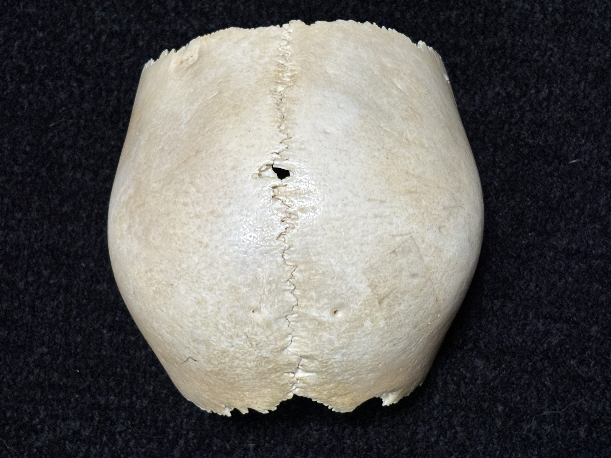
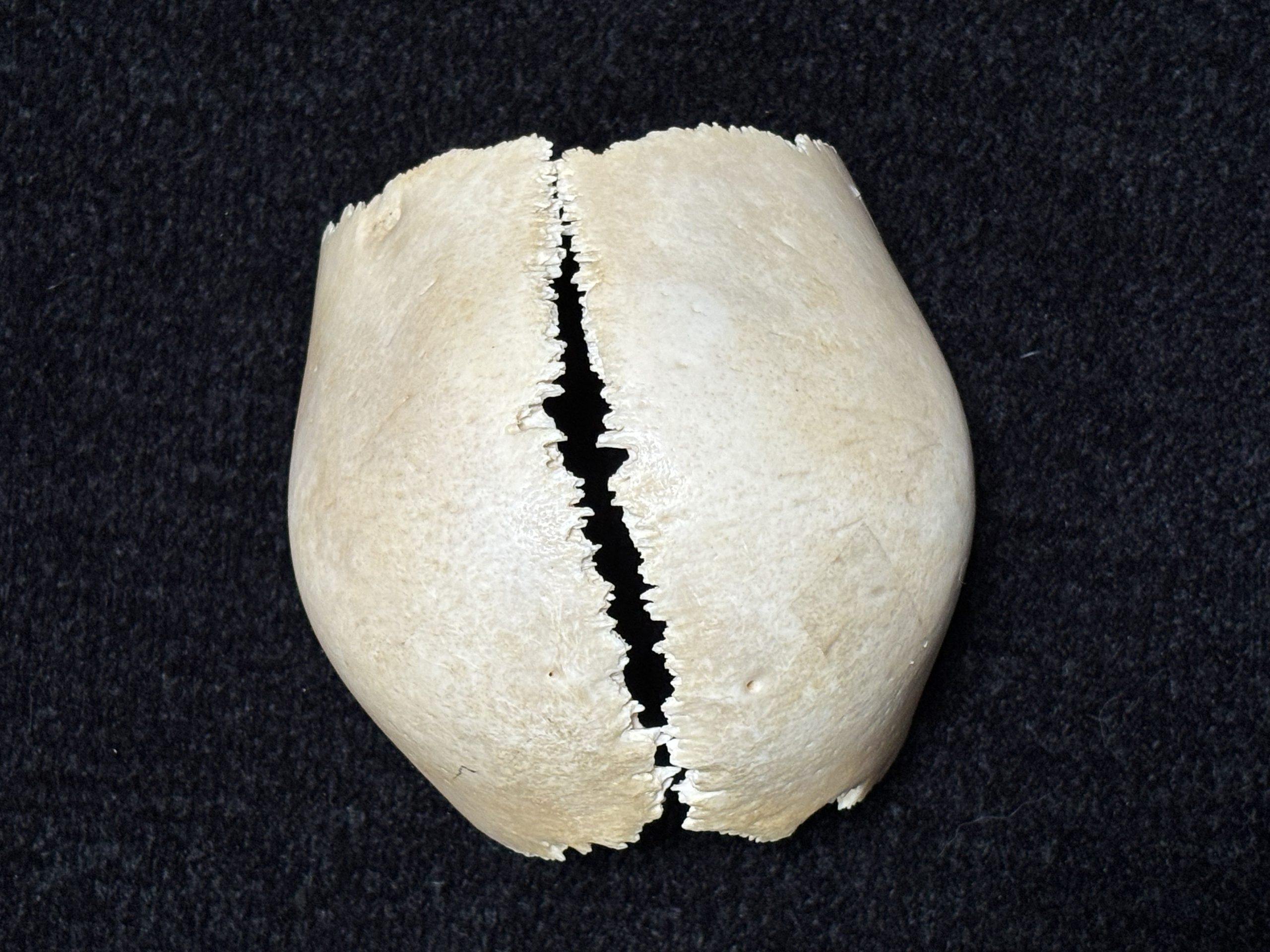
Parietal Bones | cranialconnection.com
Thanks to John McCrae, PT for specimens and photography.
The action of the tongue, the swallow and the maxillary midline suture become key players of
‘inherent cranial motion’.
The Cranial Osteopathic Orofacial Orientation adds a new dimension for both patient and doctor.
Take a look at the basic functions of life – breathing, swallowing and chewing –
from a new perspective with Tasha Turzo, DO.
TONGUE TO TOE
In the world of orofacial osteopath Tasha Turzo
– a world that encompasses an integrated outgrowth of the Swallow, the Tongue and TM Joint –
the orofacial ‘activity’ hanging from the temporal bones facilitates anterior motion of the face,
– a ‘pumping action’ for the ‘brain-drain’ of the glymphatic system.
This can only happen in the world where there is movement in the skull!
In addition, the downward suction/pulling action of the tongue (functional swallow)
on the ethmoid and vomer (the bones above the palate) at the maxillary midline suture
stimulates motion into the center of the head.
Motion in the center is exaggerated as it moves to the periphery.
I wonder if the action of the swallow and the pressure of the tongue on the maxillary suture gently expanding the palate and creating a ‘pumping action’ are the missing pieces of the glymphatic puzzle ?
…a natural way to assist the ‘physiologic motion’ of the body
to help stimulate cranial motion and drive glymphatic clearance during sleep.
The critical difference of motion vs. no motion is unmistakable from a functional perspective.
This difference is palpable, literally and figuratively.
Can there be a merger of minds and evolutionary understanding
about the Cranial Concept, the osteopathic legacy of William Garner Sutherland?
The insightful integration of cranial osteopathy with orofacial growth and development
not only directly impacts current investigations in neuroscience
but also in current practice of orthodontics and dentistry.
A Review of Glymphatics and the Impact of Osteopathic Manipulative Treatment in Alzheimer’s Disease, Concussions, and Beyond
New Discoveries with the Glymphatic System:
Pr. Maurizio de Pitta
Chikly Health Institute (2023)
Professor Maurizio de Pitta
The principal investigator of the Neuron-Glial Interactions Lab (NGILab) at the Krembil Research Institute,
Toronto, Canada.
He is an external scientific member at the Basque Center for Applied Mathematics (BCAM) in Bilbao, Spain.
Glymphatic System and
Cranial Osteopathy (2024)
Reassessing Cerebrospinal Fluid (CSF) Hydrodynamics:
A Literature Review Presenting a Novel Hypothesis
for CSF Physiology
Bruno Chikly, MD, DO
Jörgen Quaghebeur, Phd, MSc, DO
The Journal of the American Osteopathic Association (JAO)A, 2013
Is Human Cerebrospinal Fluid Reabsorbed by Lymph?
LDT and Manual Drainage of the CNS
Bruno Chikly, MD, DO
The Journal of American Academy Osteopathy
(JAAO), 1998, 8,2.
Direct Measurement
of the Rhythmic Motions of the Human Head
Identifies a Third Rhythm
Rasmussen a and Meulengracht b
The Journal of Bodywork & Movement Therapies | August 2020
a Center for Manuel Medicine, Kirke Vaerlosevj 18A, 3500, Vaerlose, Denmark
b Meulengracht Institute, Munkervaenget 26, 5230, Odense, Denmark
“The aim is to investigate the existence of a third rhythm
distinct from the head movements
caused by respiratory breathing and arterial pulsing…”
“The present study demonstrates the existence,
and normative range of a third physical rhythm
detected on the human head.”
Abstract
“Central to the osteopathic cranial field, and at the same time controversial,
is the concept of a unique rhythmic movement believed to originate
from a primary respiratory mechanism (PRM).
Further, the PRM is reported to manifest as a cranial rhythmic impulse (CRI) on the living human skull.
This study explores the rhythmic oscillations of the human head
measured directly as physical movements.
The aim is to investigate the existence of a third rhythm distinct from the head movements
caused by respiratory breathing and arterial pulsing, in an objective and purely experimental study.
This new rhythmic movement was named the primary respiratory mechanism (PRM)
by Dr. William G. Sutherland, the developer of Osteopathy in the Cranial Field
(Sutherland 1939).
Since the beginning of Osteopathy in the Cranial Field, the existence and nature of the PRM have created a continually controversial debate in scientific literature and public forums
Palpation of the CRI is central in the craniosacral evaluation
and is used worldwide by a high number of therapists as part of craniosacral assessments concerning
Cranio-Sacral Treatments (CST) and in Osteopathy in the Cranial Field.
From a scientific point of view, evidence for reliability in craniosacral assessment is not clear.”
Recording the Rate
of the
Cranial Rhythmic Impulse
Journal of the American Osteopathic Association | June 2006
ABSTRACT
“The rate of the cranial rhythmic impulse can be obtained
by both palpation and instrumentation.
However, the literature has reported higher rates obtained
by instrumentation compared with palpation.
The cranial rhythmic impulse has been demonstrated to be synchronous
with the Traube-Hering oscillation, measured in blood flow velocity.
The current study demonstrates that physicians
tend to palpate the cranial rhythmic impulse and
Traube-Hering oscillation in a 1:2 ratio
This finding provides an explanation for the difference
between palpated and instrumentally recorded rates
for the cranial rhythmic impulse.”
1:Nelson KE, Sergueff N, Glonek T. Recording the rate of the cranial rhythmic impulse.
Journal of the American Osteopathic Assoc. 2006 Jun;106(6):337-41.PMID: 16790539
Ultrasound Measurement
of Intracranial Pressure [ICP]
Magnetic Resonance Imagery [MRI] Investigates Cranial Bone Motion
Lorem ipsum dolor sit amet, consectetur adipiscing elit. Ut elit tellus, luctus nec ullamcorper mattis, pulvinar dapibus leo.