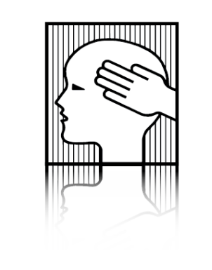
The Glymphatic System
Meets
The Cranial Rhythmic Impulse (CRI)
www.neurosciencenews.com
What is the
Glymphatic System ?
“Much remains to be studied,
but we propose
that the glymphatic/lymphatic system
acts as a cornerstone
in the architecture
of the brain and body signaling.”
The Journal of Neuroscience | 15 September 2021
What is the
Cranial Rhythmic Impulse ?
“The aim is to investigate the existence of a third rhythm
distinct from the head movements
caused by respiratory breathing and arterial pulsing…”
“The present study demonstrates the existence,
and normative range of a third physical rhythm
detected on the human head.”
The Journal of Bodywork & Movement Therapy | August 2020

Current Biology | 25 October, 2021
Lauren M. Hablitz, Maiken Nedergaard
Center for Translational Neuromedicine, University of Rochester Medical Center,
Rochester, NY 24642, USA
Center for Translational Neuromedicine,
Faculty of Health and Medical Sciences, University of Copenhagen, 2200 Copenhagen Denmark
….the brain uses a network termed
the glymphatic system
to support a constant influx of cerebrospinal fluid (CSF)
that drives the export of metabolic waste.
…to answer fundamental questions about this system…
we will need fresh perspectives, new techniques,
and collaborations across disciplines,
not only within neurobiology and sleep physiology, but also
across immunology, lymphatic biology, fluid dynamics and more.”

The Brain makes a lot of waste.
Now scientists think
they know where it goes.

A. Martinez, Host
NPR Jon Hamilton and
Jonathan Kipnis, Washington University
June 26, 2024
The Glymphatic System
Recent scientific publications illustrate the current interest in this newly discovered
Glymphatic System.
The Sleeping Brain:
Harnessing the Power
of the Glymphatic System
through Lifestyle Choices
Brain Sciences | 17 November 2020
Department of Anatomy and Neurosciences, Amsterdam
“…the glymphatic system,
which stands for glial-dependent lymphatic transport, has
been categorized as a macroscopic waste clearance system.
Due to the similarities in function,
the glymphatic system has been described as
the central nervous system’s analogue
to the lymphatic system [1,2].”
The Sleeping Brain
“Since this is a relatively new discovery,
the amount of scientific literature surrounding
the glymphatic system is rapidly increasing,
and therefore its definition is continuously being renewed.”
“This has caused controversy surrounding both the directionality
and the anatomical space in which this system resides.
Impaired glymphatic clearance has been linked to
neurodegenerative diseases [1].”
“Sleep is a primary driver of glymphatic clearance……
The physical forces propelling CSF in glymphatic clearance are intracranial pulsations.”
“Paradoxically, the N3 sleep stage which has the highest levels of CSF influx and amyloid-beta removal
also has the lowest rates of arterial pulsations [13],
suggesting that other factors are at play.
Only recently,
lower-frequency intracranial pressure oscillations
produced by respiration
were shown to complement cardiac pulsations,
which could alternatively drive clearance.”
The Glymphatic System:
A Novel Component of
Fundamental Neurobiology
The Journal of Neuroscience | 15 September 2021
Center for Translational Neuromedicine,
University of Rochester and
Center for Basic and Translations Neuroscience,
University of Copenhagen
“Much remains to be studied,
but we propose that
the glymphatic/lymphatic system acts as a cornerstone in the architecture
of the brain and body signaling.”
The Glymphatic System
“Since its elucidation in 2012,
the glymphatic system has provoked controversy,
primarily because of a lack of data
and adequate tools to characterize noninvasively
a low-pressure fluid transport system
residing in an electrically active organ
encased within the rigid walls of the skull.
Specifically, future work should focus on
how to manipulate the availability of CSF
to supply the glymphatic system,
and to elucidate the underlying
physiological mechanisms
that redistribute CSF
between the brain and the body.
The Glymphatic System, activated only during sleep,
has potential significance for neurodegenerative disease processes,
such as Parkinson’s, Alzheimer, Schizophrenia, and ALS to name a few.
As stated in the above article “The Sleeping Brain:”
“The glymphatic system is constantly filtering toxins from the brain, but during wakefulness, this system is mainly disengaged [1].
During natural sleep, levels of norepinephrine decline, leading to an expansion of the brain’s extracellular space,
which results in decreased resistance to fluid flow.
This is reflected by improved cerebrospinal fluid (CSF) infiltration along the perivascular spaces,
and therefore increased interstitial solute clearance [2].
The increase in clearance happens specifically during non-rapid eye movement sleep (N), also known as quiescent sleep.”
“Amyloid-beta and tau protein aggregations are heavily associated with Alzheimer’s disease,
creating plaques and neurofibrillary tangles in the brain that lead to brain degradation [2,3].
Glymphatic clearance moves tau proteins and amyloid-beta aggregates out of the brain [1,3].
This suggests that the glymphatic system is involved in modulating, or possibly protective against, Alzheimer’s disease.
This paper will focus on Alzheimer’s disease, since it is the most frequent dementia,
but will hopefully remain applicable to other neurodegenerative diseases, since several dementias are thought to be caused by protein aggregation.
The need for an intervention is gaining urgency [1–4].”
The Glymphatic System
Cerebrospinal Fluid Circulation
Magnetic Resonance Imaging
Provides Evidence of Glymphatic Drainage from Human Brain
to Cervical Lymph Nodes
Nature Scientific Reports | 18 May 2018
Per Kristian Eide, Svein Are Sirirud Vatnehol,
Kyrre Eeg Emblem & Geir Ringstad
“Peak CSF (cerebrospinal fluid)
tracer uptake in the
glymphatic system and
cervical lymph nodes
coincides in time.”
Eide, P.K., Vatnehol, S.A.S., Emblem, K.E. et al. Magnetic resonance imaging provides evidence of glymphatic drainage
from human brain to cervical lymph nodes.
Sci Rep 8, 7194 (2018). https://doi.org/10.1038/s41598-018-25666-4
Non-invasive MR
Imaging of Human Brain Lymphatic Networks
with Connections to Cervical Lymph Nodes
Nature Communications | 11 January 2022
Mehmet Sait Albayram, Garrett Smith, Fatih Tufan, Ibrahim Sacit Tuna,
Mehmet Bostancıklıoğlu, Michael Zile & Onder Albayram
“3D T2-Fluid Attenuated Inversion Recovery [FLAIR]
magnetic resonance imaging relies on internal signals
of protein rich lymphatic fluid rather than contrast media
and is used in the present study to visualize
the major human dural lymphatic structures.
Moreover we detect direct connections
between lymphatic fluid channels
along the cranial nerves and vascular structures
and the cervical lymph nodes..”

The Cranial Rhythmic Impulse (CRI)
https://www.headmirror.com/tbone-atlas-anatomy
The 1899 ‘AHA’ moment in medical school:
The temporal bone was
“beveled like the gills of a fish,
indicating respiratory motion
for an articular mechanism.”
Sutherland, 1967, p. 102
William Garner Sutherland
(1873 – 1954)
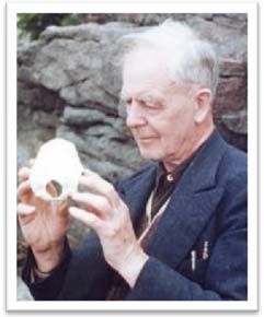
Dr. Sutherland hypothesized a
‘Primary Respiratory Mechanism’
PRIMARY
A system that comes first.
This system underlies all of life’s processes,
driving bodily functions and giving form and substance
to anatomy and physiology.
RESPIRATORY
Where does the breath originate?
A spark that gives rise to breath
that moves through tissues.
The foundation of metabolism, enabling exchange between compartments of the body.
MECHANISM
Manifesting as motion of the body,
driving the interconnectivity of the many parts
to create a functional whole that is
greater than any of the parts
functioning as separate body systems.
SIX COMPONENTS of the
PRIMARY RESPIRATORY MECHANISM
1- Cerebrospinal fluid.
2- Intracranial membranes.
3- Articular mobility of the cranial bones.
4- Spinal Cord
5- Intraspinal membranes.
6- The involuntary mobility
between the sacrum and the ilia.
The mobility between the cranial bones and
between the sacrum and the ilia
is not muscular in origin,
but rather an involuntary movement
that functions as a whole unit during respiration.
(Sutherland, 1967)
Direct Measurement
of the Rhythmic Motions of the Human Head
Identifies a Third Rhythm
Rasmussen a and Meulengracht b
The Journal of Bodywork & Movement Therapies | August 2020
a Center for Manuel Medicine, Kirke Vaerlosevj 18A, 3500, Vaerlose, Denmark
b Meulengracht Institute, Munkervaenget 26, 5230, Odense, Denmark
“The aim is to investigate the existence of a third rhythm
distinct from the head movements
caused by respiratory breathing and arterial pulsing…”
“The present study demonstrates the existence,
and normative range of a third physical rhythm
detected on the human head.”
Abstract
Central to the osteopathic cranial field, and at the same time controversial,
is the concept of a unique rhythmic movement believed to originate
from a primary respiratory mechanism (PRM).
Further, the PRM is reported to manifest as a
cranial rhythmic impulse (CRI) on the living human skull.
This study explores the rhythmic oscillations of the human head
measured directly as physical movements.
The aim is to investigate the existence of a third rhythm
distinct from the head movements
caused by respiratory breathing and arterial pulsing,
in an objective and purely experimental study.
This new rhythmic movement was
named the primary respiratory mechanism (PRM) by Dr. William G.
Sutherland, the developer of Osteopathy in the Cranial Field
(Sutherland 1939).
Since the beginning of Osteopathy in the Cranial Field,
the existence and nature of the PRM have created a continually
controversial debate in scientific literature and public forums
Palpation of the CRI is central in the craniosacral evaluation
and is used worldwide by a high number of therapists
as part of craniosacral assessments concerning
Cranio-Sacral Treatments (CST) and in
Osteopathy in the Cranial Field.
From a scientific point of view,
evidence for reliability in craniosacral assessment is not clear.”

Recording the Rate
of the
Cranial Rhythmic Impulse
Journal of the American Osteopathic Association | June 2006
ABSTRACT
“The rate of the cranial rhythmic impulse can be obtained
by both palpation and instrumentation.
However, the literature has reported higher rates obtained
by instrumentation compared with palpation.
The cranial rhythmic impulse has been demonstrated to be synchronous
with the Traube-Hering oscillation, measured in blood flow velocity.
The current study demonstrates that physicians
tend to palpate the cranial rhythmic impulse and
Traube-Hering oscillation in a 1:2 ratio
This finding provides an explanation for the difference
between palpated and instrumentally recorded rates
for the cranial rhythmic impulse.”
1:Nelson KE, Sergueff N, Glonek T. Recording the rate of the cranial rhythmic impulse.
Journal of the American Osteopathic Assoc. 2006 Jun;106(6):337-41.PMID: 16790539
Ultrasound Measurement
of Intracranial Pressure [ICP]
Magnetic Resonance Imagery [MRI] Investigates Cranial Bone Motion